Office & Emergency: 0115 989 9411

Email: info@homefarmequine.co.uk

DENTAL FACTSHEET

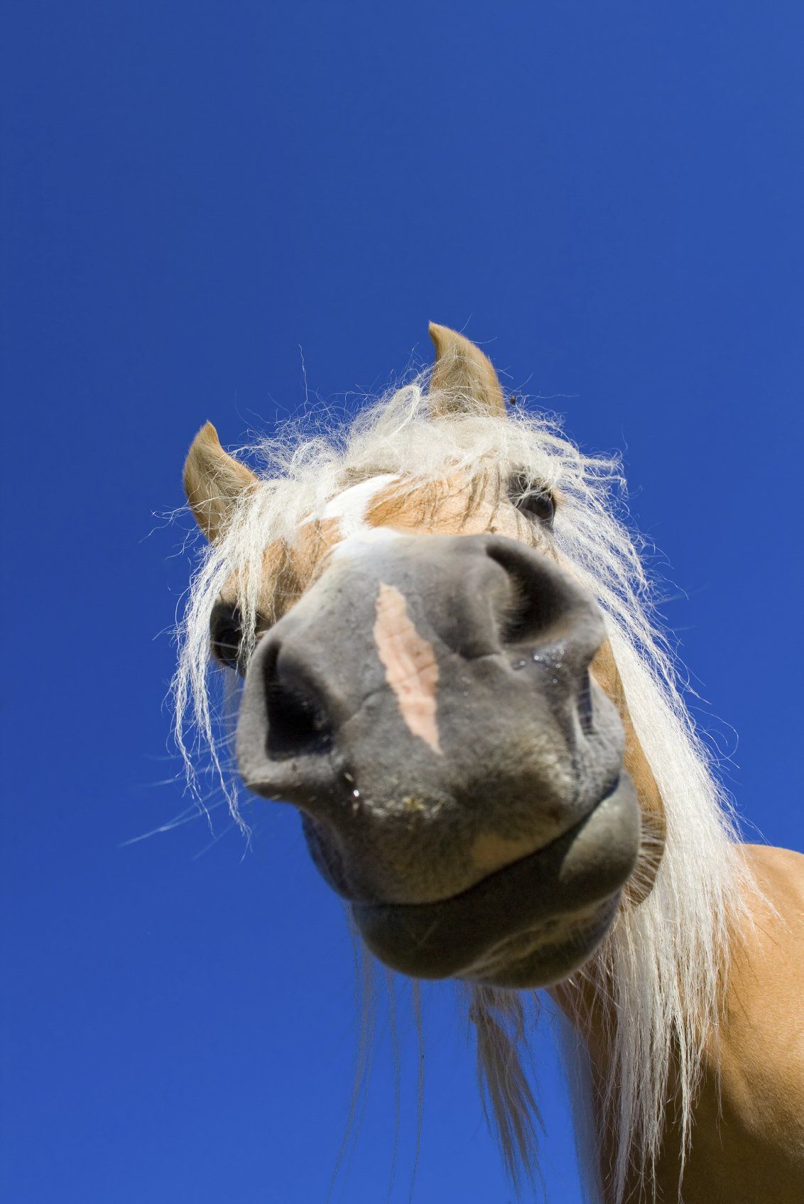
Horse’s teeth could hardly be more different than our own (see third picture above). They have 6 incisor teeth in the upper and lower jaws and then a large gap before the six cheek teeth. We use the term ‘cheek teeth’ to describe the premolar and molar teeth collectively. Male horses also have canine teeth which emerge at about 5 years of age in the gap between the incisors and molar teeth in both upper and lower jaws. They are only occasionally seen in mares.


Incisor and Canine Teeth
Problems with the incisor and canine teeth are thankfully rare and are mainly the result of trauma, such as kicks from other field-mates or hooking their teeth on a hay net.
Some geldings develop tartar and calculus on their canine teeth and require this to be cracked off each year. Unless they develop cavities like their sweet toothed owners! Incisor teeth start to wear out in the mid to late 20’s but this is variable.


Cheek Teeth
These are termed hypsodontal which means that, unlike ours, they do not have a distinct crown above the gum and root below. 
The constant grinding action of a horse chewing wears away the emerging tooth. In the wild the rough forage and sand picked up with grazing means this process of emergence versus wear and tear remains in fine balance.
However, with a nicely manicured pasture and soft palatable concentrate feeds, the teeth don’t wear away as fast as they would in the wild and so develop overgrowths of enamel at the grinding edges of the upper and lower cheek teeth.


Routine Dental Care
Your horse should have its teeth checked annually by an equine vet or an accredited Equine Dental Technician (EDT). It is important that you take the time to ensure the person you are trusting with your horse’s teeth is a properly qualified. ANYBODY can call themselves an ‘equine dentist’ with absolutely no training or qualification.
The vast majority of routine dental care is to address this issue of enamel overgrowths, which develop into sharp points (below left) or enamel transverse ridges (ETR below right).

If left untreated the commonest signs of enamel overgrowths are:
  • Quidding - dropping food.
  • Balling – food pouches in the cheeks like a hamster
  • Inappetence - reluctance to eat
  • Tossing of the head or biting discomfort.
  • Smaller enamel overgrowths can be easily removed with a hand rasp or ‘float’. A gag must be placed in your horse’s mouth to safely and properly inspect and rasp the teeth. About 1 in 20 horses find this frightening and require light sedation. The way to avoid this is to have your horse checked each year from two years old so that they are used to having a ‘Gag’ put into their mouth. Most horses do not need any actual rasping until they are 4- or 5-year-olds when they have all their adult cheek teeth in place.
  • Some horses will need these enamel overgrowths removed twice yearly. More severe overgrowths – hooks and ramps (see below) - do occur and are often caused by malformation of the teeth such that they do not have an opposing tooth to wear against.
  • Hooks and ramps require motorised burrs to remove these large areas of diamond-hard enamel. They commonly recur at the same rate that the tooth is erupting, i.e., 2-3 mm per year, and so require treatment every year or so


Diastema

Diastema are caused by malformation of the teeth so that food jams between two adjacent cheek teeth. Daily grinding packs more and more food into the tiny space, eventually forcing food under the gum line. Diastema can be difficult to identify, often requiring the use of a dental mirror. The picture on the left below shows food jammed at the ‘6 o’clock’ position in the mirror.

Diastema are a common cause of oral pain and require specialist treatment to either widen the space between the two teeth, allowing the food to move freely in and out of the gap, or to extract the cheek tooth if it has become diseased.

It is important to identify diastema early as widening the space can save the tooth. The gap is widened using a special motorised burr (above right). This procedure may require repeating as the teeth grow and the gap closes.


Cheek Teeth Fractures and Abscesses

Fractures and abscesses involving the cheek teeth invariably require extraction of the offending tooth. This is a major undertaking in horses due to the massive length of the ‘root’.

Traditionally teeth were extracted by making a hole in the face (for an upper tooth) or the jaw (for a lower tooth) and hammering out the tooth. Now it is much more common to extract teeth from the mouth by using large dental extractors (below) to loosen a diseased tooth and pull it free.

Oral extractions require substantial patience as it can take 6-8 hours to wiggle the tooth loose. Great care must be taken not to fracture the tooth or leave fragments behind. The teeth at the bottom left of the picture above are from a 20-year-old horse and only a third of the length of those in 7 year-old.

Occasionally, and especially in young horses, it is not possible to extract a tooth orally. In these cases, the horse is anaesthetised and a dental punch inserted through a hole made in the jaw bone or face to hammer out the tooth. The picture on the right above shows an Xray taken during surgery to check that the dental punch is correctly positioned prior to being hammered.


Wolf Teeth

About a third of horses have wolf teeth that erupt just in front of the cheek teeth between 1 and 8 years of age. They serve no purpose and are a remnant of the first pre-molar tooth that is no longer required. About 50% of horses need to have it removed as it interferes with the bit. This is easily done under light sedation.

There are many areas not covered in this fact sheet, but we hope it has helped you understand your horse’s mouth. Horses have quite amazing teeth and a simple check-up at vaccination time should keep them that way.


If you have any further questions, please do not hesitate to contact us on

0115 989 9411
.


by Home Farm Equine 06 Oct, 2021
SYMPTOMS The typical signs of strangles include: • Sudden high temperature and loss of appetite. • Cough. • Slight yellowy nasal discharge. • Swollen lymph nodes (glands) under the jaw and in the throat: never present in the early stages of the disease. • Rupture of the abscesses in the affected lymph nodes. Horses usually recovered fully and naturally after the rupture of the abscesses. Once the horse has recovered from strangles it usually develops a strong immunity and it is rare for an animal to be affected more than once. If your horse shows any of these symptoms it is important that you contact your vet to help confirm the diagnosis as soon as possible. A blood test and/or nasal swab can be taken to determine if you horse does have strangles. We will then be able to guide and help you with the treatment and management of your horse for their quicker recovery and also to prevent an outbreak of the disease. DIAGNOSIS The time between infection and development of clinical signs varies but it is usually 1 to 2 weeks. Strangles is diagnosed primarily on the clinical signs which may be strongly suggestive of its involvement. Nasal swabs samples can be taken for laboratory investigation. Recently a blood test has been developed which is able to tell us whether the horse has had contact with strangles in the recent past. A negative result for strangles in the swab or the blood do not guarantee that the horse does not have strangles, but these laboratory results together with the symptoms can be very helpful in determining if the horse is affected and which horses may be involved when an outbreak occurs in the yard. Some horses can become carriers, where they do not suffer the disease but they can spread it to other horses.
by Home Farm Equine 06 Oct, 2021
THE EXAMINATION The standard examination is conducted in either two or five stages, although the exact sequence of the examination may vary. If a two stage is required a disclaimer form will need to be signed and returned previous to the vet attending. The stages are: Stage 1: Preliminary examination This is a thorough external examination of the animal at rest using visual observation, palpation and manipulation to detect clinically apparent signs of injury, disease or physical abnormality. It includes an examination of the incisor teeth, a thorough examination of the horse's eyes in a darkened area and auscultation of the horse's heart and lungs at rest. Examination of the eyes does not include dilating the pupil but should include examination of internal and external structures. The examination DOES NOT include examination of the inside of the prepuce (sheath), a detailed mouth examination with a speculum, a height measurement or any examination for pregnancy. Stage 2: Walk and trot, in hand The animal is walked and then trotted in hand to detect abnormalities of gait and action. Ideally this is carried out on firm, level ground. The horse is turned sharply each way and is backed for a few paces. Flexion tests of all four limbs and trotting in a circle on a firm surface may be carried out if the examining veterinary surgeon considers it safe and appropriate to do so. Stage 3: Exercise phase The horse is usually ridden and given sufficient exercise to: Allow assessment of the horse when it has an increased breathing effort and an increased heart rate. 2. Allow assessment of the horse's gait at walk, trot, canter and, if appropriate, gallop. 3. Allow assessment of the horse for the purpose of stage five. If ridden exercise is not possible for any reason then this stage may be conducted by exercising the horse on a lunge, but this fact should be made clear to the purchaser and on the certificate. Stage 4: Period of rest and re-examination The horse is allowed to stand quietly for a period. During this time the respiratory and cardiovascular systems may be monitored as they return to their resting levels. Stage 5: Second trot up The animal is trotted in hand again to look for any signs of strains or injuries made evident by the exercise and rest stages. Flexion tests Flexion tests and trotting in a circle Flexion tests and trotting in a circle on a firm surface are not mandatory parts of the standard procedure, but they can sometimes provide useful additional information about a horse. There may be circumstances when the examining veterinary surgeon concludes that it is unsafe or inappropriate to perform such tests. Blood Sample Blood Sample A blood sample may be taken for storage (usually for 6 months) for possible future analysis to detect substances present in the horse's system at the time of the examination that might have masked any factors affecting the horse's suitability for the purchaser's intended use. If a blood sample is not taken then the reason should be noted on the certificate. Identification Identification of the horse The horse should be identified by recording the horse's markings in the form of a diagram and written description as well as searching for a microchip and inspecting any available documentation. The diagram may be omitted if the presence of a microchip can be confirmed by scanner and the diagram of an accompanying passport matches the horse. In this instance, both microchip and passport numbers should be recorded on the certificate.
by Home Farm Equine 06 Oct, 2021
As the temperatures begin to rise and the sun starts to shine, regrettably so does the risk presented to the horses caused by Sycamore seedlings and seeds. Eating these can cause Seasonal Pasture Myopathy (previously known as Aytypical Myopathy / Sycamore poisoning). This condition has a very sudden in onset, with clinical signs consisting of: stiffness, reluctance to move, muscle tremors, sweating, weakness, lethargy, difficulty in breathing, reluctance to work, red or brown urine, choke, colic, low head carriage, fast or irregular heartbeat and sudden collapse. The time from the onset of symptoms to collapse can be as short as 24 hours. The helicopter shaped seeds are now springing up as seedlings, which are also poisonous. We would advise pulling up all seedlings before allowing horses to graze any affected pasture. Seeds can travel up to 200metres from the tree, especially in the winds we have been experiencing lately, so please be vigilant. Seedlings, seeds and leaves of Sycamore trees can be tested for the toxin Hypoglycin A, which causes SPM. The Royal Veterinary College, London is carrying out this testing. http://www.rvc.ac.uk/research/laboratories/comparative-neuromuscular-diseases-laboratory/diagnostic-services#panel-n-e-w-atypical-myopathy-testing If your horse shows any of these signs, or you are worried after grazing near sycamore trees, please call us immediately on 0115 9899411 .
by Home Farm Equine 06 Oct, 2021
When to castrate? Most horses are castrated as yearlings, however anytime after 6months is acceptable providing both testicles have descended. When colts mature into stallions, and they reach 2 years old, the testicles become much larger and have bigger blood vessels which make the castration procedure more difficult as they require ligation (tying off) of these vessels. It is normal to try and avoid the fly season from May to October as the castration wounds are left open and do attract flies which is a major cause of post castration infection. How to castrate? Well-handled colts up to 18months old can usually be castrated standing with heavy sedation. However, if they are likely to kick or have unusually large testicles they will need to be “knocked down” and given a short general anaesthetic (GA) which makes the process safer for all concerned and allows the vet to tie off the large testicular blood vessels. The process usually takes 15 minutes and then a further 15-30 minutes for the horse to stand up again. An area of flat, clean grass or a school surface is required. Post castration care Routine care is simple but essential and without these complications are very common. The castration site wounds are always left open to allow the post–op swelling fluid to drain away. The removal of the testicles leaves a cavity which has to fill in and heal from the inside; this process produces inflammatory fluid which must be allowed to drain. It is ESSENTIAL that the wound is hosed TWICE daily for 5 minutes and a scab, if present, is wiped away (with some cotton wool in dilute Savlon or Hibiscrub) to stop the scrotal skin edges sticking together and trying to heal and close. The wound must be kept open for at least a week. Failure to do this will allow the wound to close too soon and a large fluid swelling at the site and swelling of the sheath will occur. This will then require the vet to visit and reopen the site and put the horse on a course of antibiotics. It is very important that the horse is turned out to EXERCISE as this will reduce the amount of swelling of the sheath. Even with exercise it is normal for the sheath to swell to half a honey melon size. We normally recommend that the colts are turned out the same day as the castration, but adult stallions are turned out the day after castrate. Tetanus cover is essential and if not vaccinated it will need an anti toxin injection to give immediate protection. Pain relief is recommended with “bute” given twice daily for 5-7 days. Antibiotics are not routinely given as these surgeries are left open to drain and thus are not required. Although castrations are considered to be a very routine procedure it must be remembered that in the horse they are a relatively major surgical operation with potential fatal complications such as haemorrhage and evisceration of small intestine through the surgical wound. These complications can be reduced to almost “theoretical” providing the correct techniques are used in relation to the size, age and type of male horse being treated. For owners the mainstay of care is thorough cold hosing and cleaning of the wound for at least a week and providing good clean turnout exercise. Contact with mares should be avoided for one month. If you have any further questions, please do not hesitate to contact us on 0115 989 9411.
by Home Farm Equine 06 Oct, 2021
The following information will cover points on the following: Wounds Colic Lameness Choke WOUNDS LOCATION OF THE WOUND Wounds on the leg sheath/joints on the fore and hind legs need an examination. The horse may be acutely lame with a tendon sheath/joint infection. THIS IS A TRUE EMERGENCY AND A VET MUST BE CALLED AS SOON AS POSSIBLE
by Home Farm Equine 06 Oct, 2021
Veterinary Acupuncture is a complementary treatment that is increasingly being recognised as a useful treatment for a wide range of conditions. It should be stressed that the term “complementary” is the correct term for the use of veterinary acupuncture, as this therapy complement our conventional veterinary care. It is an adjunct, not a replacement, although in some situations it can be used as a sole treatment.
by Home Farm Equine 06 Oct, 2021
What is Cushing Disease? The average age of horses with Equine Cushing disease is 19 years old with a range of 7 to 40 years. There is no sex predisposition but ponies appear to develop the disease more frequently.
by Home Farm Equine 06 Oct, 2021
Parasite Control in Horses Horses can become infected with both ectoparasites (lice and mites) and endoparasites (worms). The effect of a parasite infection on the horse's health depends on the number and type of parasite involved. Generally, the higher the parasite burden, the higher the risk of clinical disease. 
More posts
Share by: